Med Spa
A Med Spa service is a non-surgical cosmetic (or hair removal) service performed under the supervision of a licensed medical doctor. Medical spa services comply with HIPAA regulations, so your information is kept confidential. Medical spa services require more training in technique and safety than regular spa services, and can often be performed by a doctor, physician assistant, or a licensed aesthetician.
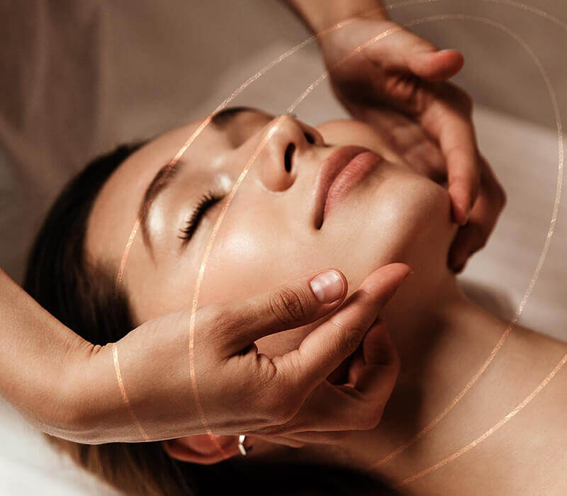
Treatments
Medspa treatments cover a wide range of services, including skin care product recommendations, Botox, dermal fillers, laser hair removal, Broadband Light therapy (BBL/IPL), and micro laser peels. Many medspas carry medical-grade skin care products that are not available anywhere but in select doctor’s offices. These products are stronger, more concentrated, and more effective than your usual drugstore or makeup counter skin care products.
Med Spa In Virginia
- Botox
- BroadBand Light Therapy (BBL/IPL)
- Chemical Peels
- Dermal Fillers
- Facials
- Laser Resurfacing
- Laser Hair Removal
- HydraFacial Treatment
- NanoLaserPeel
- Laser Scar Revision
- Microneedling
- PRF Treatments, including Hair Restoration and Facial Rejuvenation
NOVA Plastic Surgery offers all the above-mentioned medspa services with our highly trained and skilled Physician Assistants, Michele Gittings, PA-C and Taylor Poe, PA-C. We also carry SkinCeuticals, Silagen scar cream, and Latisse for purchase.
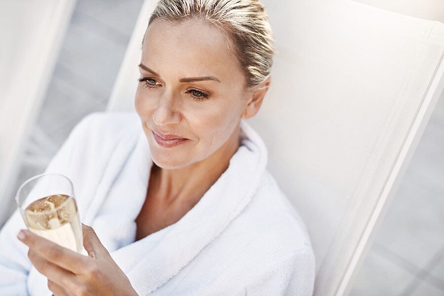
Most of the medspa services we offer can be done on your lunch break (or in a similar short time period), and have little-to-no downtime, so you can resume your regular activities as soon as you leave.
Some of the services, such as laser hair removal or Broadband Light Therapy, require several sessions spaced out over a few weeks in order to be most effective.
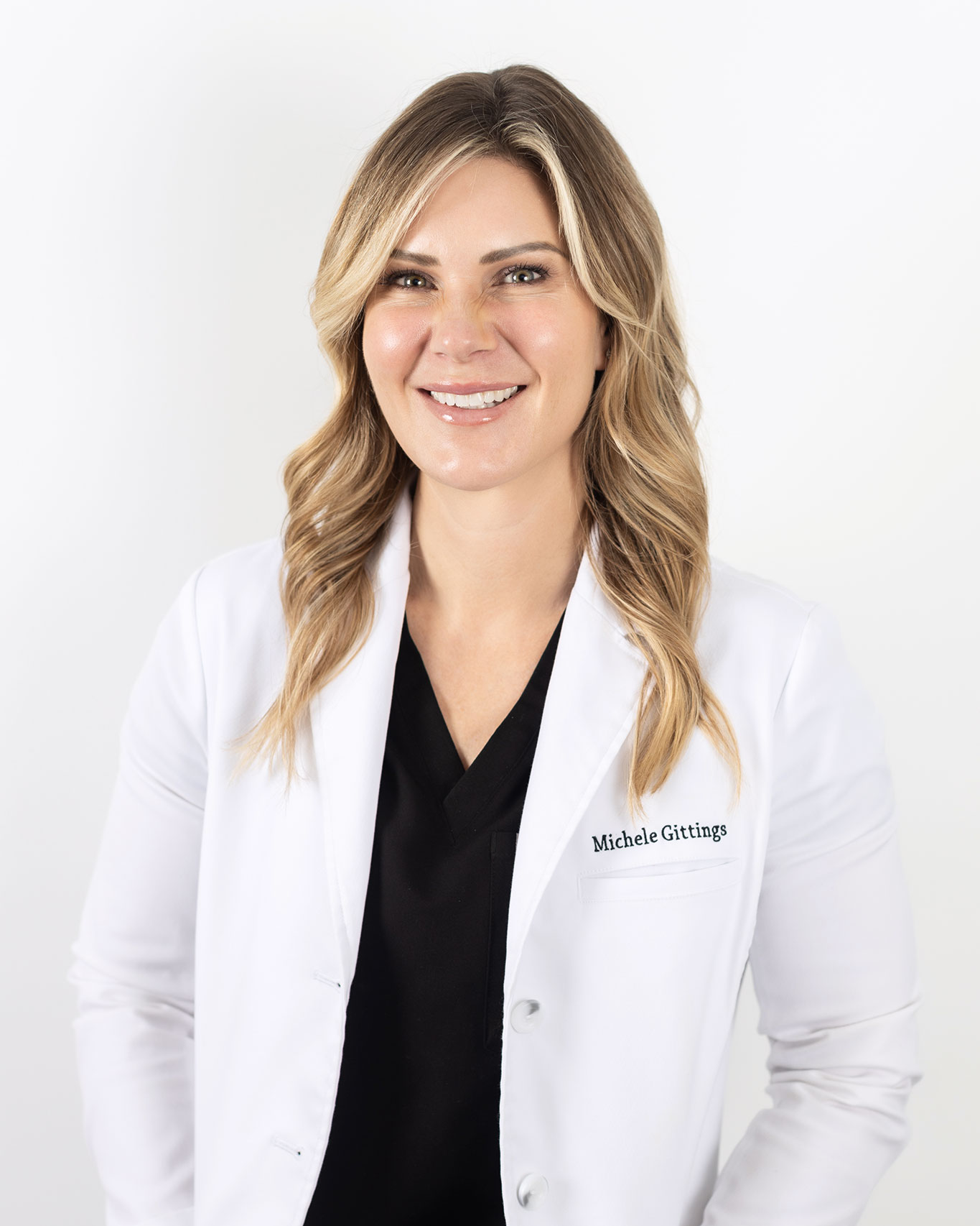
Michele Gittings
Michele Gittings is a Nationally Board Certified Physician Assistant who received her degree from St. Francis University in Loretto, Pennsylvania. Michele is an experienced and talented aesthetic physician assistant. Michele evaluates and treats the wide spectrum of clients with facial rejuvenation concerns. She is an expert on providing all our medspa services especially our signature non invasive, Liquid Facelift, that will make you look younger and more refreshed without the need for plastic surgery.
Learn More About MicheleTaylor Poe
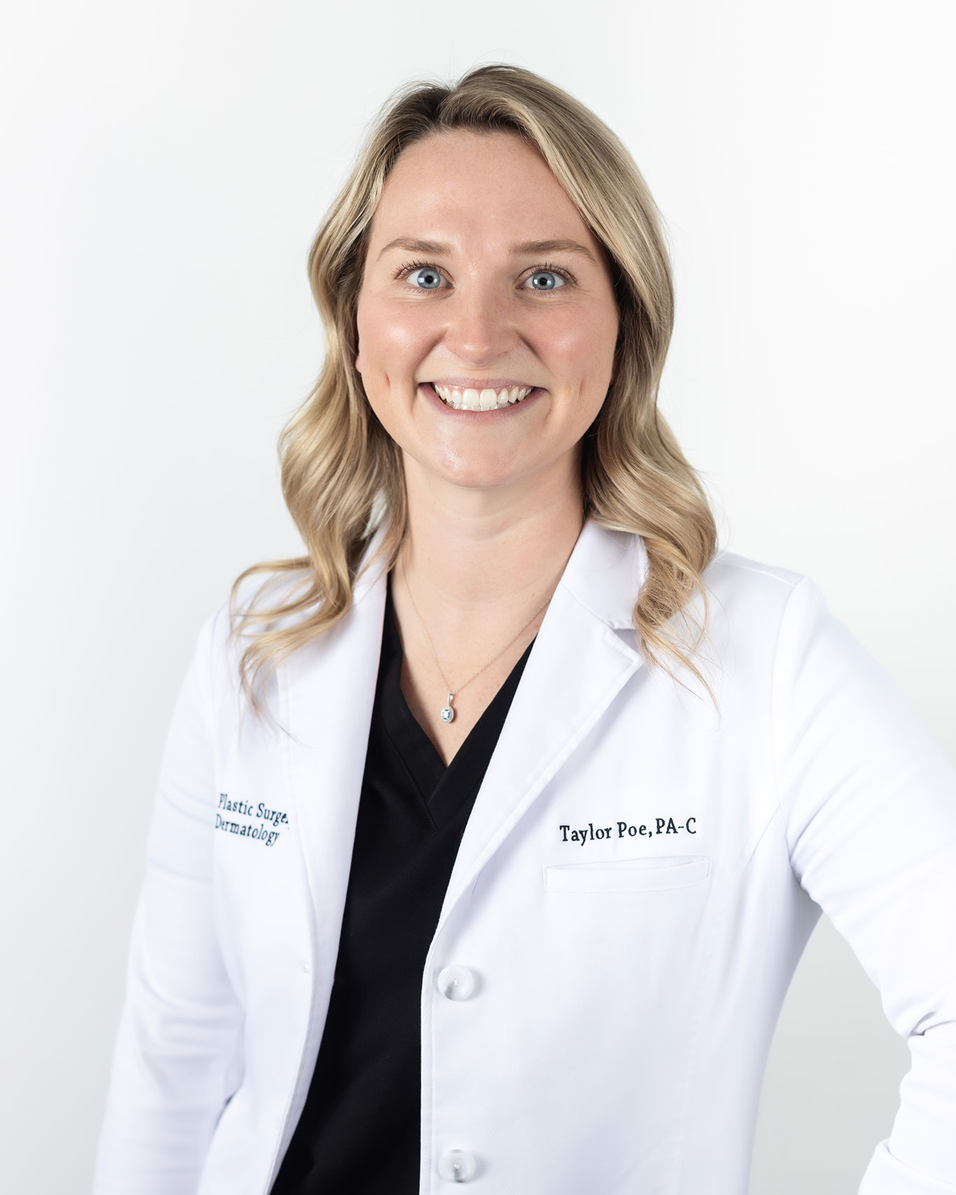
Taylor Poe is a Nationally Board-Certified Physician Assistant who received her Bachelor’s Degree from Virginia Tech then continued onward to receive her Master’s Degree in Physician Assistant Studies from East Carolina University where she finished top of her class. She evaluates and treats a wide spectrum of cosmetic and skin concerns of the face and body. Along with assisting Dr. Nukta in surgery, she is highly skilled in the use of non-invasive facial rejuvenation treatments including botox, fillers, and skin treatments.
Learn More About Taylor